UvA Master study Cell Biology & Advanced Microscopy (BMS-CBAM)
Introduction
Cell Biology is the discipline that studies the function of cells in the complexity of tissues and organs in the human body in order to understand mechanisms of disease. The tremendous revolutions in the past decade in light microscopy and biosensors to visualize processes in cells have changed cell biology completely.
Cell Biology and Advanced Microscopy (CBAM) is a track within the 2-years Biomedical Sciences Master (BMS) study and a collaborative effort by the University of Amsterdam (UvA) the Academic Medical Centre (AMC) and the Netherlands Cancer Institute (NKI), who collaborate in the van Leeuwenhoek Center for Advanced Microscopy (LCAM). These departments are all front runners in studying cell functions microscopically in time, even down to the single-molecule level.
The objectives
The major objective of this track is the understanding of fundamental cell biological research in relation to human disease. This understanding comprises:
1. cell biology in the context of the healthy and diseased human body;
2. the cellular basis of major human diseases such as neurodegenerative diseases, infection by viruses, bacteria and parasites, and cancer such as leukemia;
3. diagnostics such as cytopathology and prenatal screening & fertility-related disorders; and
4. advanced microscopy including live cell imaging, correlative light and electron microscopy, confocal microscopy and the underlying biophysics.
To visualize processes in the cell at the microscope fluorescent proteins will be used. By recombinant DNA technologies
you can optimize the fluorescent proteins, biosensors that for example can monitor intracellular calcium or protein interactions,
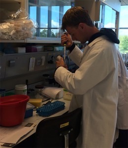
or couple your favorite protein of interest to a fluorescent label. CBAM student Dave Huiskens created new constructs for better fluorescent proteins.
Programme Outline
The programme consists of three core courses and the participation in two or three internship research projects (9 or 6 months). The research projects lead to the writing of a thesis manuscript in English that is in principle suitable for publication in a scientific journal. Furthermore, two optional specialized courses in human disease such as cytopathology, medical microbiology, hematological diseases, fertility & sterility will be offered.
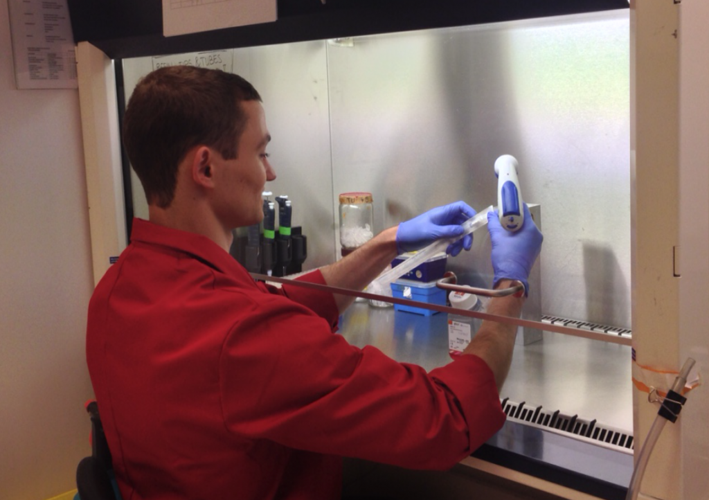 Experiments are performed in single cell or cell organoids cultures. One has to passage the cell lines
in order to keep them alive and introduce the recombinant DNA by transfection protocols into the cells
after which the cells will start producing the fluorescently labeled proteins themselves.
Core courses
▪ The first course is Molecular Biology of the Cell. This basic theoretical course of 4 weeks that is the same for all master tracks in the Medical Biology cluster will recap the molecular basis of cell function including proteins, membranes, DNA structure, cell metabolism, protein synthesis and cellular signaling.
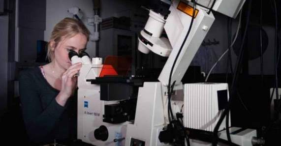
▪ The second course is Advanced Microscopy. The aim of this 4-weeks practical course is to get hands-on experience and theoretical background in performing state-of-the-art (light) microscopy applied to cell biology. The microscope setups on which the training is provided, are part of the van Leeuwenhoek Centre for Advanced Microscopy (LCAM) in Amsterdam. During the first three weeks every day you will start with a lecture discussing a different fluorescence microscopy technique followed by a tutorial session. In the afternoon you will sit at the dedicated microscope and do the experiments yourself. In the final week you will select and perform a mini-research project in one of the three LCAM laboratories where you can apply and strenghten the acquired knowledge of the previous weeks.
Movie of AnexinA4-GFP protein dynamics in transfected HeLa cells using confocal microscopy
as acquired by CBAM students Cristiaan Wessels, Christiaan Zeelenberg and Tomasz Gora
during the mini-research project week of the Advanced Microscopy course 2011.
▪ The third course is Clinical Cell Biology. This theoretical course of 4 weeks starts with a general lecture course treating cellular signaling, the cytoskeleton, mechanism/regulation of cell division, structure of cells in tissues, apoptosis, fertility, cellular basis of cancer and metastasis, infection by viruses. The majority of the course consists of actual clinical cell biology topics of research groups at the UvA, AMC and NKI. Topics will include: Structure/function of invapodia, mechanisms of cancer cell migration, role of TRPM7 channels & signaling (NKI); Neurodegenerative diseases, viral/bacterial infection & cytopathology, DNA-damage/repair mechanisms in relation to cancer (AMC); GPCR-signalling, cell division machinery, Ca2+ signaling, signalosome formation at membranes, biosensors and pharmacology (UvA).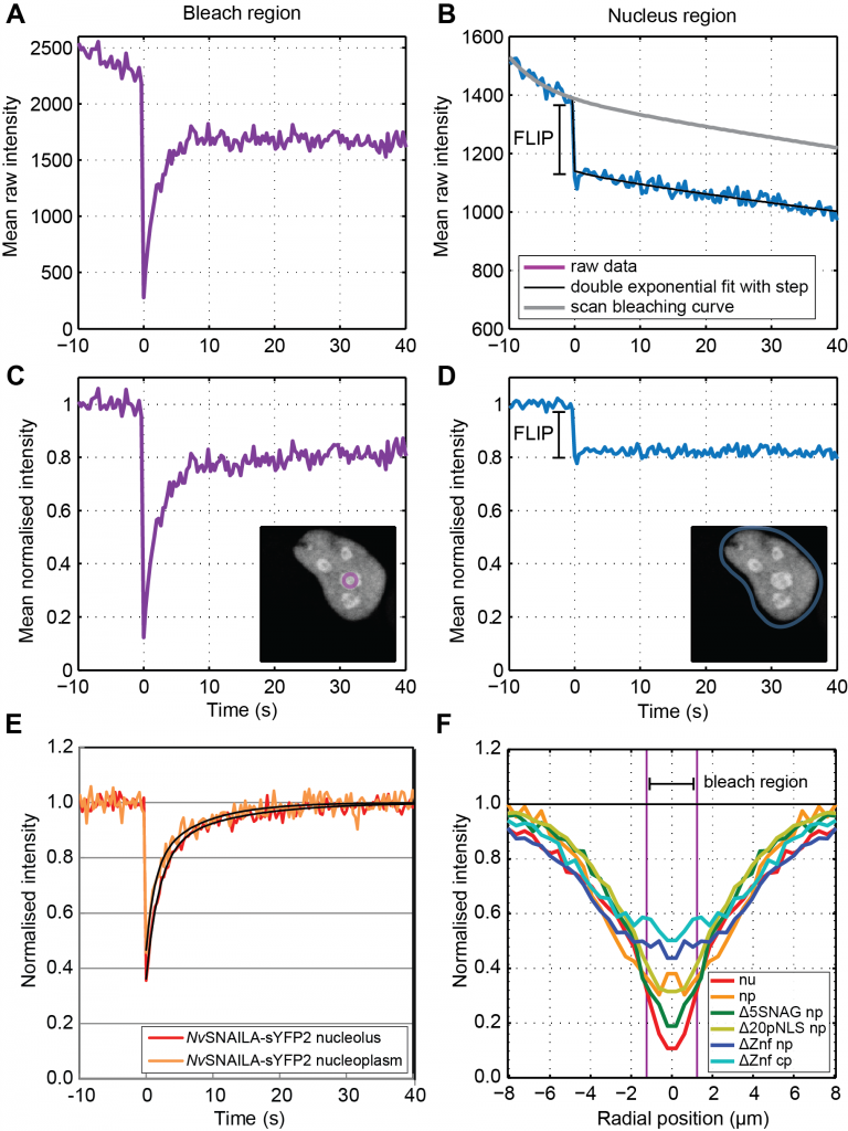
After the images have been collected the data has to be analysed. You will have to select the appropriate datasets, fitting model, if wished write your own
analysis-software and interpret the results. Besides the localisation of the protein the advanced microscopy techniques nowadays allow you to get much more information
out of your images like concentration, transport speed or the degree of interaction with other proteins.
Here the mobility of nuclear protein SNAIL is monitored at various sub-cellular locations via Matlab scripts (image from Marten Postma & Ada Dattoli).
Internship research projects
Advanced microscopy applied to clinical cell biology problems will form the backbone for the research projects. The LCAM partners at UvA, AMC and NKI provide the research projects in close collaboration with scientists in which the master students can participate. A typical example of such a research project is to create your own genetic encoded sensor (using GFP labeling and cloning techniques), cell transfection & manipulation, performing experiments with living cells using advanced microscopes, interpreting image data, image analysis and processing and finally the design of follow up experiments. It is strongly advised to do at least one research project at one of the three participating departments in LCAM and one research project abroad in one of the many international research labs that collaborate with LCAM.
Is Cell Biology & Advanced Microscopy the programme for me?
The programme is looking for students who:
• are interested in the molecular and cellular mechanisms underlying disease
• are excited about visualizing cells in action using live cell microscopy
• have an interest to learn state-of-the-art single cell microscopy
• are creative and want to design their own genetically encoded biosensors to probe molecular disorders responsible for diseases such as cancer, infection, sterility and problems related to aging
• enjoy labwork and working in a dynamic research team using sophisticated microscopical techniques
• may want to develop novel state-of-the-art image analysis software or microscopy driver software (optional)
• want to persue a scientific career in top life sciences research departments
 Click here for an interview (Dutch) with CBAM student Nathalie Reinhard during
her internship research project at the section of Molecular Cytology
(published in Folia Magazine, 4 April 2012, foto:Jan-Maarten Hupkes)
For more info about this Master study click here for the UvA website or contact the track coordinators Dorus Gadella (FNWI) or Eric Reits (AMC). The UvA organizes on regular basis “meeloopdagen” where you can join a current CBAM student for a day, to get an impression how the Masterstudy Cell Biology & Advanced Microscopy will look like.